Introduction
Long-chain n-3 polyunsaturated fatty acids (PUFAs) play crucial roles in various physiological processes. Docosahexaenoic acid [DHA, 22:6(n−3)] is particularly essential for proper development and function of the brain and retina throughout life. The molecular structures of dietary lipid molecules delivering the n-3 PUFA likely influence the digestion and absorption of these valuable fatty acids (Yoshinaga et al., 2015). Currently, there is very limited knowledge on the regio- and stereospecific position of n-3 PUFAs in dietary lipids affects their bioavailability and metabolism.
Objectives
The objective is to improve the current understanding of the significance of positional distribution in TAG molecules for the bioavailability and metabolic fate of DHA. To reach this objective, we performed an animal feeding trial was performed and studied the FA composition in plasma, liver, brain, and visceral fat of rats after intervention feeding with regio- and enantiopure triacylglycerols (TAG) containing DHA in sn-1, 2, or 3 position and palmitic acid (16:0) in the remaining positions (Figure 1).
Study Design
Seventy-two male Sprague-Dawley rats (age 21 ± 2 days) were kept for 7 days in isolation at a constant temperature and humidity and on an adaptive feeding of the standard AIN-93G diet which contained soybean oil as a source of (n-3) FA. Thereafter, the rats were randomly divided into six experimental groups, 12 animals in each group receiving normal feed, n-3 deficient feed, or n-3 deficient feed supplemented with tripalmitate or DHAcontaining TAGs (Table 1). In DHA groups (3-6), each rat received the structured TAG at a daily dosage of 500 mg /kg body weight, and the intervention feeding lasted for four weeks. At the end of feeding, the rats were executed after 12 hours of fasting, plasma and organs were collected and stored at -80 C. The lipids were extracted using the modified Folch method (Folch et al., 1957), and the fatty acids were analysed as FAMEs by gas chromatography (Linderborg et al., 2019). For plasma and liver, the lipids were fractionated into neutral lipid fraction rich in TAGs and polar lipid fraction rich in phospholipids (PL), thereafter, fatty acids were analysed from these fractions.
Results
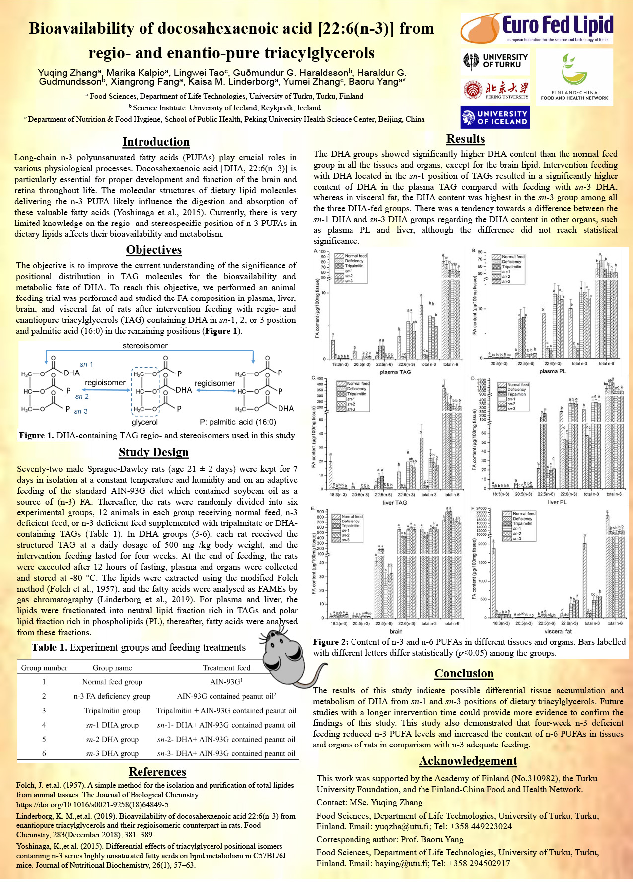
The DHA groups showed significantly higher DHA content than the normal feed group in all the tissues and organs, except for the brain lipid. Intervention feeding with DHA located in the sn-1 position of TAGs resulted in a significantly higher content of DHA in the plasma TAG compared with feeding with sn-3 DHA, whereas in visceral fat, the DHA content was highest in the sn-3 group among all the three DHA-fed groups. There was a tendency towards a difference between the sn-1 DHA and sn-3 DHA groups regarding the DHA content in other organs, such as plasma PL and liver, although the difference did not reach statistical significance.
Conclusion
The results of this study indicate possible differential tissue accumulation and metabolism of DHA from sn-1 and sn-3 positions of dietary triacylglycerols. Future studies with a longer intervention time could provide more evidence to confirm the findings of this study. This study also demonstrated that four-week n-3 deficient feeding reduced n-3 PUFA levels and increased the content of n-6 PUFAs in tissues and organs of rats in comparison with n-3 adequate feeding.
References
Folch, J. et.al. (1957). A simple method for the isolation and purification of total lipides from animal tissues. The Journal of Biological Chemistry. https://doi.org/10.1016/s0021-9258(18)64849-5 Linderborg, K. M.,et.al. (2019). Bioavailability of docosahexaenoic acid 22:6(n-3) from enantiopure triacylglycerols and their regioisomeric counterpart in rats. Food Chemistry, 283 (December 2018), 381–389. Yoshinaga, K.,et.al. (2015). Differential effects of triacylglycerol positional isomers containing n-3 series highly unsaturated fatty acids on lipid metabolism in C57BL/6J mice. Journal of Nutritional Biochemistry, 26(1), 57–63.
Acknowledgement
This work was supported by the Academy of Finland (No.310982), the Turku University Foundation, and the Finland-China Food and Health Network.
Contact: MSc. Yuqing Zhang
Food Sciences, Department of Life Technologies, University of Turku, Turku, Finland. Email: yuqzha@utu.fi; Tel: +358 449223024
Corresponding author: Prof. Baoru Yang
Food Sciences, Department of Life Technologies, University of Turku, Turku, Finland. Email: baying@utu.fi; Tel: +358 294502917
